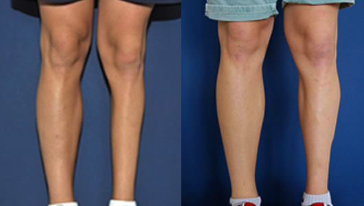
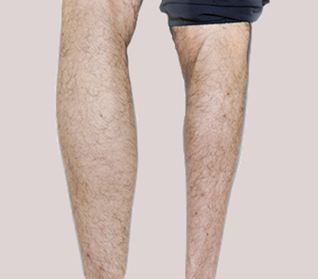
Back view of the calf atrophy of the right leg

How to treat calf atrophy ?

Other cosmetic defects regarding soft tissues can also be managed with custom-made implants, particularly the calf atrophy

As the implants’ designing corresponds perfectly to the patient’s anatomy, many cases can be considered to correct congenital or acquired muscular hypotrophies or ageneses, due to :

  • a talipes equinovarus
  • the sequelae of a polyomyelitis
  • degenerative diseases
  • traumatic and/or surgical sequelae


Body

Definition and origin of the calf atrophy

Atrophy of the calf corresponds to a lower volume of the leg (unilateral or bilateral), and concerns mainly the muscle groups of the posterior part (gastrocnemius), incidentally on the knee, leg and ankle bones.

The source of this atrophy can be innate or constitutional. Acquired atrophies occurs after a disease or a surgery : poliomyelitis, clubfoot, amyotrophic lateral sclerosis, lupus, meningocele, foot surgery. There is a deformity, which is often unilateral, and an asymmetry compared to the other limb.

The functional consequences are generally moderate or due to restricted physical activities induced by poor body image. The objective of this correction is purely morphological.

3D custom-made technique

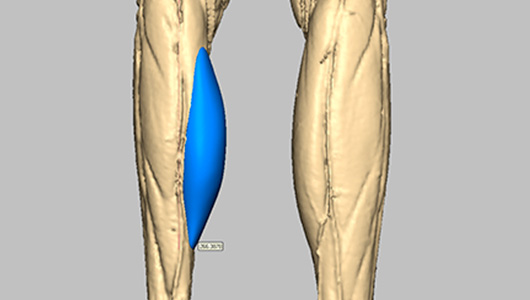
From a 3D scan of the patient, we make a virtual copy of their calf, including each tissue (bone, muscle, skin, cartilage). The implant is then virtually designed by computer and the prototype machined as a perfect copy of the implant. Our partner Sebbin takes care of the final manufacturing of the silicone implant from this prototype, following the ISO standards. This technique enable us to obtain a definitive implant, with invisible edges, perfectly adapted to the anatomy of the patient's calf.

Learn more

Surgery and follow-up

Calf atrophy surgery with 3D custom-made implant

Surgery

  • It concerns the technique created and published by Dr Glicenstein in 1975, updated by Pr Chavoin with computer designed silicone gum implants.
  • The surgery takes place en prone position, under general anaesthesia and lasts less then 1 hour and presents minimal risks.
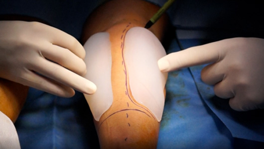
  • The surgeon traces the location of the lodge, makes an incision in the hollow of the knee and places the implant. The duration of hospitalisation is 3 days at most.
  • 1 to 3 days hospitalisation maximum.

3D view of the custom-made implant targeting calf atrophy surgery

Surgical outcomes

  • Surgical outcomes with little pain: the pain is treated with simple analgesics
  • No post-operative care: the stitches are resorbable, no drain, only a dressing for 8 days
  • Progressive standing up and walking are prescribed activities as soon as the next day to avoid the risk of thrombosis and related to anticoagulants. Compression stockings are not necessary
  • There is no major sero-hematic or serous effusion
  • Sick leave for 15 days and sports restriction for 3 months

Results

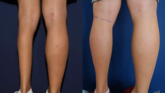
If this technique brings a real improvement regarding the volume, it cannot offer a perfect symmetry for atrophies of both the skeleton and the joints. The distal third of the leg, lacking muscle, is imperfectly corrected by the implant and might justify, subsequently, an additional transplant of fat tissues.

Vor der Wadenatrophie Operation Ergebnis

Ergebnis einer Wadenatrophieoperation

Testimonials

Surgeon

An important part of my work is also calf augmentation or reconstruction. And custom made computer design implants are extremely good in achieving as much symmetry as possible between the 2 legs.

Dr Tillo, London (United Kingdom)

Patient

I had an operation at the Regina Maria Pia Studio to increase the volume of my right calf, because I was born with a club foot and therefore a slightly atrophied calf. I did various research and decided to trust DR.

Lorenzo, 24 years old, Roma