Clinical Scenario
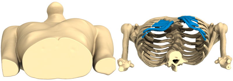
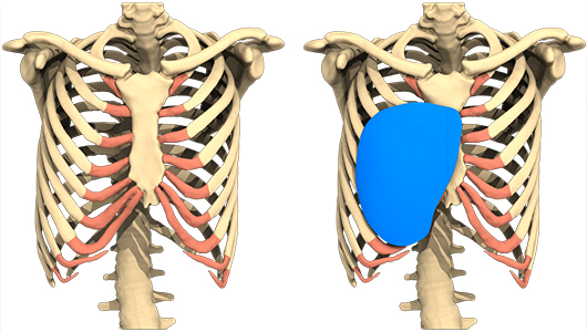
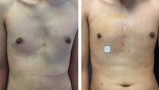
He is a 28-year-old man who came to my consultation for a chest deformity problem with a sagging aspect on the right side. This deformation is congenital, but has worsened over time since childhood. The diagnosis of pectus excavatum is worn, it is a pectus type 3, according to the Chin classification, completely lateralized to the right and with a projection of the left costal medial awning. He does not complain of any respiratory disorder.
We explain the possibility of correction by setting up a custom 3D implant. A detailed information sheet is given to him on his pathology and his treatment with a custom implant. Before making a decision, we prescribe a chest CT scan and a cardio-respiratory assessment (EFR, VO2max, ecg, echocardio) in principle.
A second consultation confirms the good results of the functional exploration (VO2max 67% theo = 26.6 ml / min / kg), the scanner confirms the diagnosis and the absence of associated malformations. The custom implant is ordered and the intervention scheduled three months later.

The surgery
The operation is done under general anaesthesia, the large implant is placed by a 8-cm-long vertical medial approach in front of the sternum, after marking on the skin of the precise area of its compartment. The dissection is done under the pectoral muscle and the fascia of the right abdominal muscle, it extends quite low under the external oblique muscle, which is unusual. The intervention time is 50 minutes. After closing the muscular and cutaneous planes.
The outcomes are simple with the wearing of a compression bra and analgesic treatment with paracetamol. The sero-thematic collection is punctuated on D1, D2, D7, and D14.

Result
The result is excellent for the patient and the left costal projection is very attenuated by the adjacent filling. The patient keeps his compression vest for one month day and night. He can return to work after 1 month and to the sport gradually after the control consultation at three months.
The patient does not request the correction of his prominent cartilage, which has completely disappeared with the implant filling.

Author and bibliography
Author
Dr Debrosse is a thoracic and pulmonary surgeon with more than 20 years of experience this field at the Tenon hospital in Paris and formerly at the Institut Mutualiste Montsouris. He has also been a volunteer for the Franco-Vietnamese Association of Pneumology.


